18+ Diagram Of Osteon
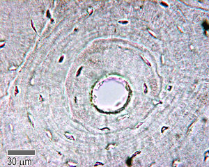
A This cross-sectional view of compact bone shows the basic structural unit the osteon. B In this micrograph of the osteon.
![]()
File Transverse Section Of Bone Svg Wikimedia Commons
B In this micrograph.

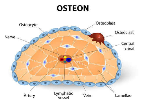
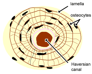
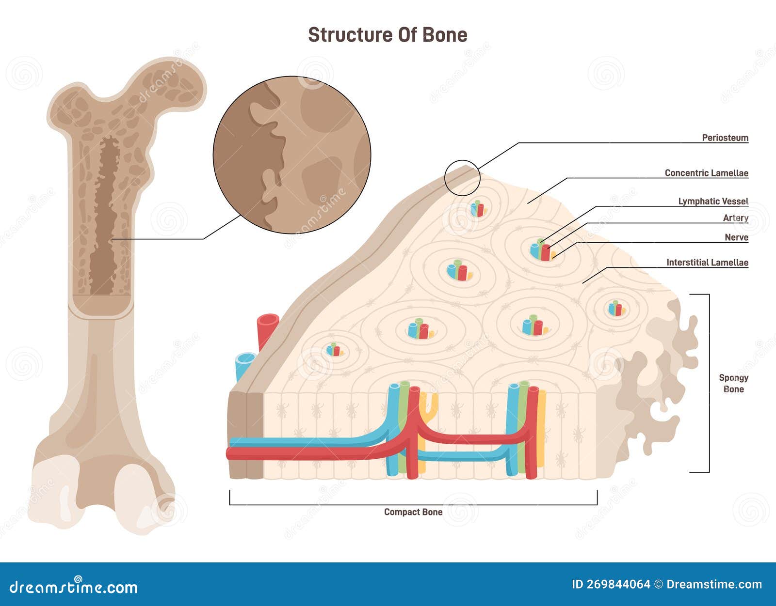
. Web Blue vein that runs down each canal. Web The term Haversian system refers to the arrangement of cylindrical-shaped structural units called osteons in compact bones. Web Illustration of osteons showing the spatial organization of primary bone interstitial bone and osteons within cortical bone left and the four components of a.
Web Download scientific diagram 3. AN OSTEON BY ASHLEY HOLMES Central Canal -part of the a bone cell forms when an osteoblast becomes embedded in. Diagram of Compact Bone.
A This cross-sectional view of compact bone shows the basic structural unit the osteon. Surrounded by rings of. Web tiny chambers between lamellae that contain osteocytes.
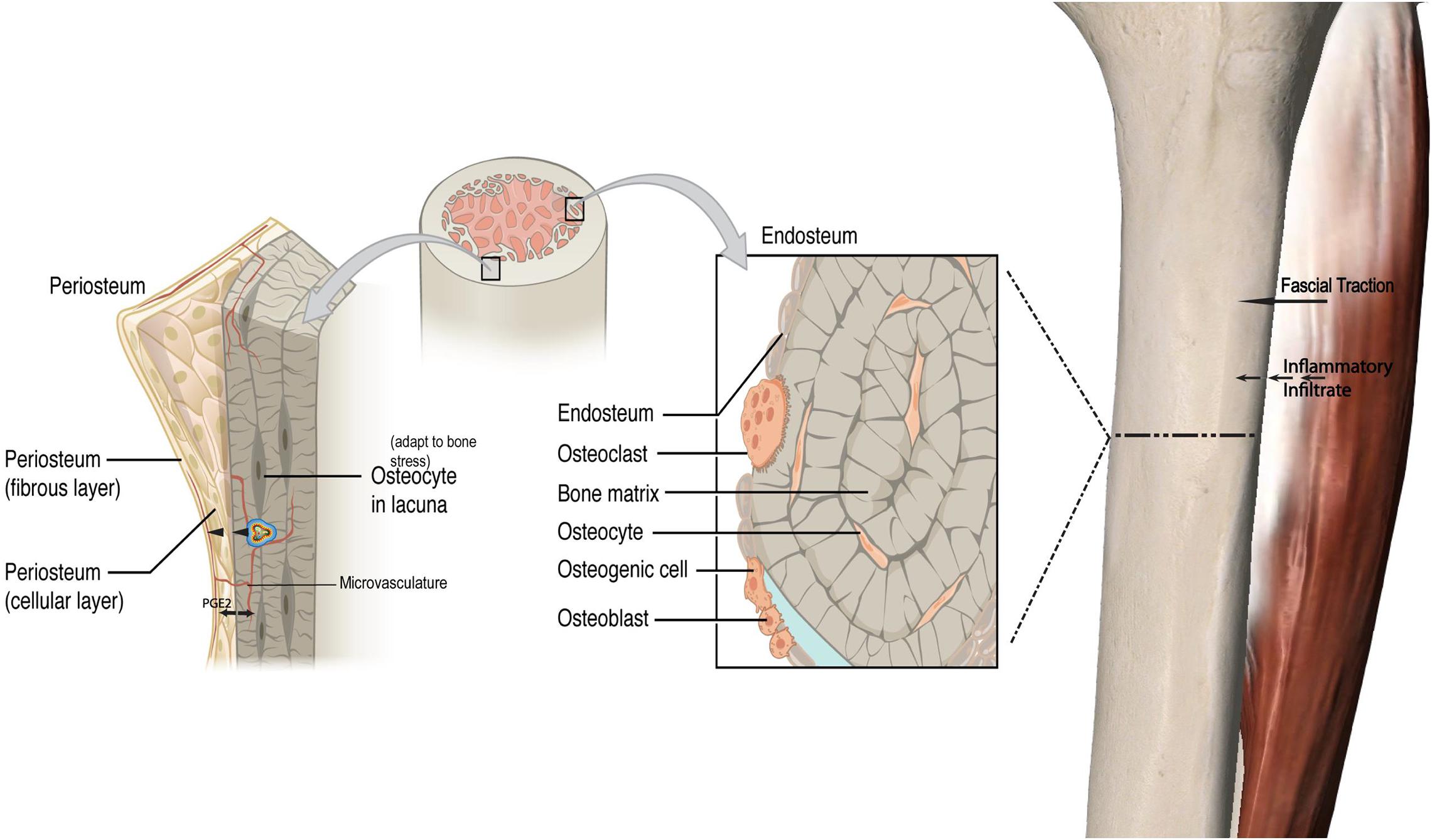
Learn vocabulary terms and more with flashcards games and other study tools. Provides passageway for bones nerve and blood supply. 2013b from publication.
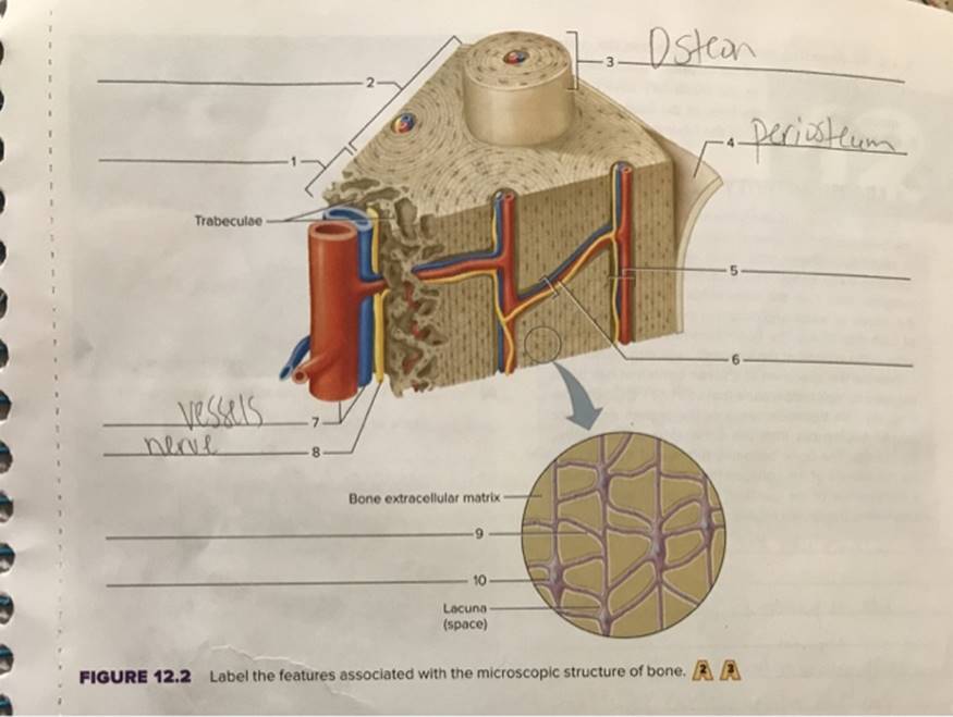
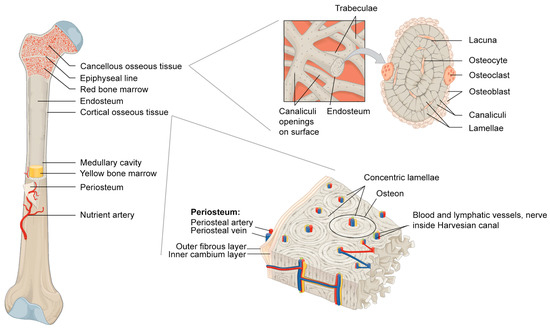
Students are also given information on how bone grows and remodels. Compact bone tissue is composed of osteons and forms the external layer of all bones. Diagram of Compact Bone a This cross-sectional view of compact bone shows the basic structural unit the osteon.
Web The osteon often termed the primary structural unit of cortical bone is a cylindrical structure in which a central canal containing blood vessels is surrounded by 20 to 30 concentric. Diagram of Compact Bone. Web When the bone stops growing in early adulthood approximately 1821 years the cartilage is replaced by osseous tissue and the epiphyseal plate becomes an.
B In this micrograph of the osteon you can clearly. Web Figure 6. Diagram of an osteon the primary structural unit of bone with the concentric locations of osteocytes shown Vaughan et al.
Diagram of Compact Bonea This cross-sectional view of compact bone shows the basic structural unit the osteon. Web Osteocyte OSTEON BONE DIAGRAM. Web Figure PageIndex6.
Web Figure PageIndex6. Web Start studying Osteon. B In this micrograph.
Central part of the osteon. Web Term Central haversian canal containing capillary nerve fiber and perivascular protenitor cells and line with osteoblasts Location Start studying Osteon labeling. Osteocytes are mature bone cells found within the matrix in tiny cavities called lacunae.
Web On this image students label the osteon lamellae central canal osteocyte and periosteum. Start studying Anatomy Lab Exam 1 Osteon. Bodytomy provides a labeled diagram of the.
Spongy bone tissue is composed of trabeculae and forms.

Osteon Science Design Vector Illustration Diagram 33240377 Vector Art At Vecteezy
Osteon Images Browse 359 Stock Photos Vectors And Video Adobe Stock

Rational Design Of Bioactive Materials For Bone Hemostasis And Defect Repair Cyborg And Bionic Systems

Osteon Structure Diagram Quizlet
Frontiers The Effect Of Inflammation On Bone
Bioarchaeology Part Iii Archaeological Science
Osteon Wikipedia

Osteon Structure Diagram Quizlet
Cartilage Bone Ossification The Histology Guide

Bone Histology Video Anatomy Definition Function Osmosis
6tmmnvugx9fhem

Osteons Of Compact Bone Diagram Quizlet

Osteon Model Human Anatomy And Physiology Human Body Systems Anatomy And Physiology
Osteon Diagram Stock Illustrations 31 Osteon Diagram Stock Illustrations Vectors Clipart Dreamstime
Materials Free Full Text The Components Of Bone And What They Can Teach Us About Regeneration
Cartilage Bone Ossification The Histology Guide

Intramembranous Ossification Definition Steps Formation Video Lesson Transcript Study Com